
Simultaneous fluorescence optical and electrical characterization of atrial tissue
- Ansprechperson:
- Projektgruppe:
Digitale Signalverarbeitung
Cardiac arrhythmias are among the leading causes of death in the Western world. The gold standard for the treatment of cardiac arrhythmias, like atrial fibrillation, is radio frequency ablation (RFA). The aim of RFA is to find focal triggers or modified substrate areas causing the arrhythmia using multipole catheter electrodes inside the heart measuring intracardiac electrograms. Depending on the type of arrhythmia several ablation procedures are used. However, no reliable or robust criteria to assess lesion formation, e.g. linear lesions, exist up to the present time and the long-term success of catheter ablation is not guaranteed.
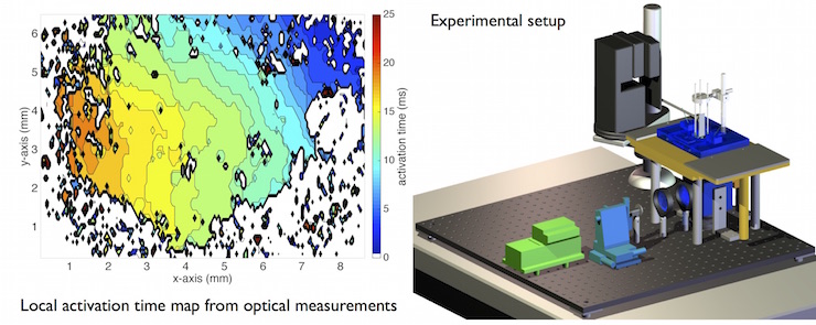
At the Institute of Biomedical Engineering in Karlsruhe, an in-vitro measurement setup was established measuring the electrical activity of vital cardiac preparations under defined conditions. The propagation of the cardiac excitation is monitored with fluorescence-optical techniques by using a voltage sensitive fluorescent dye (Di-4-ANEPPS). Extracellular potentials are recorded simultaneously with a miniaturized electrode. The main objective of this research project is to investigate the electrophysiological characteristics of cardiac tissue with acutely created ablation lesions. Therefore, the temporal development of the extracellular potentials on the cardiac tissue will be measured with a multielectrode array after the ablation procedure. Furthermore, an optical beat to beat acquisition of excitation patterns surrounding ablation lesions will be feasible with high spatio-temporal resolution.
An acute ablation lesion and its border zones are described in a current computational model by various physiological parameters changing the extracellular signals temporarily or permanently. The further development of this computational model allows to simulate complex ablation patterns and to determine the electrograms around these lesions. Electrophysiological studies as well as ablation procedures are performed at the Städtisches Klinikum Karlsruhe. The intracardiac electrograms are recorded permanently during the electrophysiological examinations. The clinical results will be compared with simulation studies as well as experimental results from the in-vitro setup.
Publications
- M. Keller, S. Schuler, et al., “Characterization of radiofrequency ablation lesion development based on simulated and measured intracardiac electrograms,” IEEE Trans Biomed Eng, vol. 61, pp. 2467–2478, 2014.
- M. Keller, Formation of Intracardiac Electrograms under Physiological and Pathological Conditions. PhD thesis, KIT Scientific Publishing, 2014