
Cardiac Magnetic Resonance Image Segmentation
- Typ:Studentische Forschungsarbeit
- Betreuung:
Hiwi Stelle
-
Motivation and Background
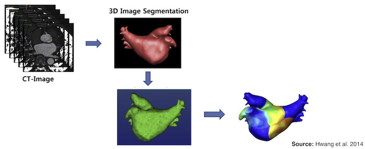
Computational modeling is a field that is continuously growing and personalized models need patients' data. Several efforts have been done in this direction, but most of them require a big amount of human interaction, which is both time consuming and could be a source of error. Late gadolinium enhancement (LGE) magnetic resonance imaging (MRI) segmentation provides the atrial anatomy as endocardial and epicardial surfaces with an approximate distribution of fibrotic tissue.Fibrosis modifies the patients' substrate where the action potential propagates creating the perfect conditions to initiate and/or maintain an arrhythmia. Unrecognized or left untreated arrhythmias can cause life-threatening complications that may affect the heart and/or brain. However, it is still unclear how the information of the LGE-MRI images of the atrium can be used to characterize the patients' substrate. In this project, the student should manually segment LGE-MRI images obtaining patients specific geometries and proposed an algorithm for a semiautomatic segmentation.
Project
This project will bring you the opportunity to learn about cardiac anatomy, clinical, MRI images and segmentation tools used in different fields. The main objective of this project is to manually segment LGE-MRI images, obtaining the endocardial and epicardial surfaces as well as the fibrotic region surface. Furthermore, a semiautomatic segmentation algorithm should be proposed.Skills
- Good programming skills (Python)
- Ability to work independently
- Commitment and self-organization skills
- Communication and work will be done in English