
Spatial-temporal Analysis of Multichannel Electrocardiogram with special Focus on P-wave
- Forschungsthema:Spatial-temporal Analysis of Multichannel Electrocardiogram with special Focus on P-wave
- Typ:Diplomarbeit
- Betreuung:
- Bearbeitung:
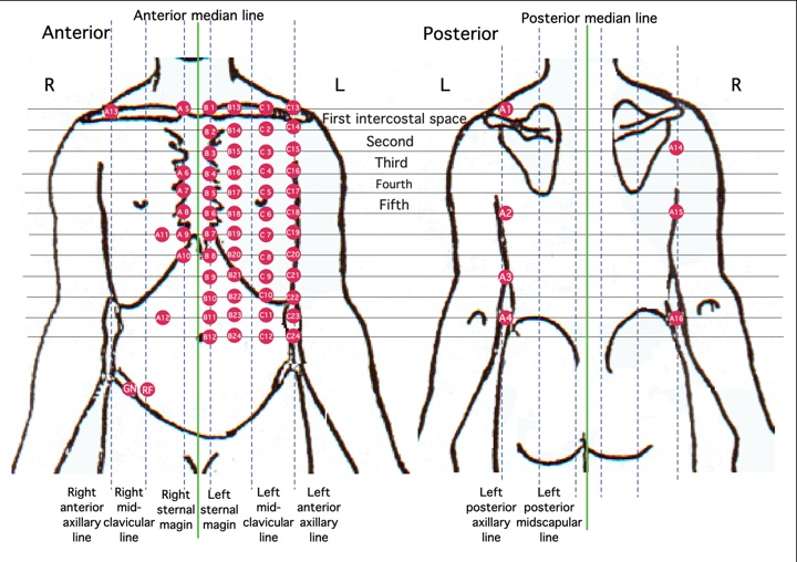
The ECG is a bioelectric signal, which records the heartís electrical activity versus time, therefore it is an important diagnostic tool for assessing heart function. The Standard 12-Lead Electrocardiography gives a measure of cardiac activity with high temporal resolution. Multi-channel ECG (64 leads) is an extension of the conventional 12-Lead ECG which gives temporal as well as spatial resolution.
Different parts of the ECG give different diagnostic information. The morphology, amplitude and time of occurrence of the P-wave has got lot of significance, due to its diagnostic importance in the case of atrial abnormalities. Multi-channel ECG measurement and analysis is performed to detect the small changes in P-wave morphology temporally and spatio-temporally as well as to examine the relationship between the morphology changes and heart rate.
This diploma thesis is focused on the measurement and analysis of Multi-channel-ECGs. Therefor measurements at rest and under stress conditions of healthy volunteers should be performed. From the measured data the P-waves should be extracted and then be analysed by Principal Component Analysis. The results will be shown in a Body Surface Potential Map as graphical presentation of cardiac activity as measured on the surface of the human torso.
 |
 |
| 64-Channel-ECG Electrode Positioning | Body Surface Potential Plot of p-Wave activity |