
Estimation of Conduction Velocities and Excitation Patterns in Optical Mapping Data Using Ratiometry
- Typ:Bachelorarbeit
- Betreuung:
- Bearbeitung:
-
Motivation
At the Institute of Biomedical Engineering an experimental setup was established measuring the electrical activity of vital cardiac preparations under defined conditions. Besides the electrical recording of the extracellular potentials, the propagation of cardiac excitation is measured with fluorescence-optical techniques. In this project, we are going to investigate the electrical activity of living myocardium with acute ablation lesions. In a next step, a new High-speed camera has been integrated in the in-vitro setup to detect the signal of a voltage sensitive fluorrescent dye (di-4-ANEPPS) with high spatial-temporal resolution over a longer time period.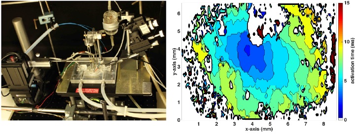
Project description
In a first experiment, the electrical activity of the right atrium of rat myocardium was recorded with this new High-Speed camera. Aim of this thesis is the analysis of the optical mapping data before, during and after the radiofrequency ablation. For this purpose, it is required to load the raw data and to select manually specific time periods in a GUI. By adapting the process parameters, the extracted image data will be analysed to estimate local activation times, excitation patterns as well as conduction velocities in different regions of the myocardium. This program offers a reproducible and efficient data evaluation.