
Detailed Anatomical and Electrophysiological Modeling of the Human Ventricle based on Diffusion Tensor MRI Data
- Forschungsthema:Detailed Anatomical and Electrophysiological Modeling of the Human Ventricle based on Diffusion Tensor MRI Data
- Typ:Diplomarbeit
- Betreuung:
- Bearbeitung:Dipl.-Ing. David Keller
Diplomarbeit
Detailed Anatomical and Electrophysiological Modeling of the Human Ventricle based on Diffusion Tensor MRI Data
Diplomarbeiter: David Keller
Betreuer: Dr.-Ing. Gunnar Seemann, Dipl.-Ing. Daniel Weiß
Zeitraum: 01.02.2006 - 31.07.2006
Betreuer: Dr.-Ing. Gunnar Seemann, Dipl.-Ing. Daniel Weiß
Zeitraum: 01.02.2006 - 31.07.2006
Simulation of the electrical and mechanical properties in the human ventricles requires a precise knowledge of structure and electrophysiology. On one hand, electrophysiological properties are known to be heterogeneously distributed within the ventricles. On the other hand, the structure of the ventricles has an oriented and laminated structure influencing electrical conduction and deformation.
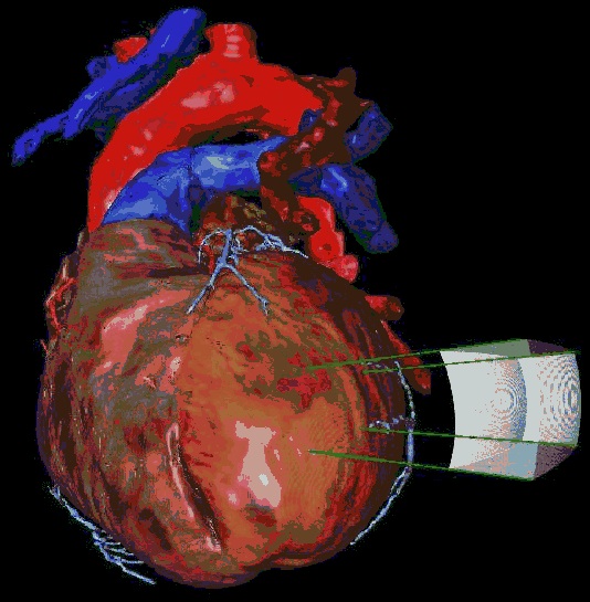
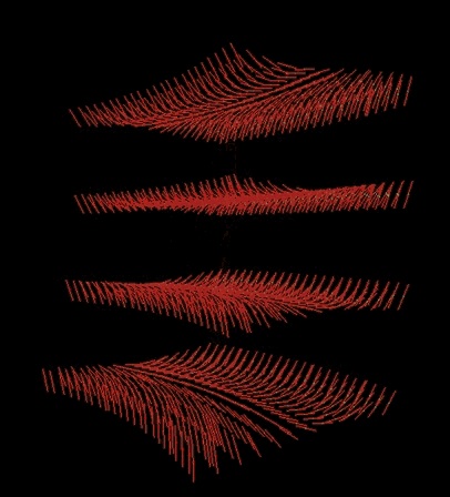
This work focuses on generating a realistically electrophysiological and anatomical model of the human ventricles based on Diffusion Tensor (DT) MRI data provided by Raimond Winslow of the Johns Hopkins University, Baltimore. Therefore, the ventricles are segmented with active shape and interactive deformable meshes in left and right ventricle including e.g. papillary muscles. The orientation of the muscle fibers is extracted from the DTMRI data and is assigned to the geometrical model. From that data, rules are extracted to define the fiber orientation within different locations of both ventricles. These rules are compared to the data of Streeter and other fiber orientation data.
The electrophysiological model of ten Tusscher et al. is applied in conjunction with the bidomain model to the geometrical model. Knowledge of transmural electrophysiological heterogeneity is assigned to each region and the physiological excitation is computed. Furthermore, the electrophysiological model is adapted to long QT syndrome type and the effects of this mutation are investigated.