Robust conduction velocity estimation in a clinical setting
- Typ:Masterarbeit
- Betreuung:
- Bearbeitung:
-
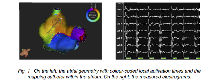
Motivation
Atrial flutter (AFlut) is a supraventricular tachycardia which affects 1.2% of hospitalised patients. This disfunction of the human atria goes hand in hand with an increased risk of stroke and other life-threatening diseases. The therapy of AFlut is a standard procedure. Electrophysiologists monitor the excitation origin and propagation on the endocardial surface of the atria during a minimally invasive procedure. The electroanatomical mapping system automatically processes the measured electrograms. The activation times of the atrial tissue that is in contact with the measurement electrodes are the basis of the physician’s diagnosis. Activation times are colour-coded and visualised on the individual atrial geometry. Regions of slowed excitation propagation are assessed as arrhythmogenic, i.e. a part of the pathological excitation pathway. After identifying them, the physician tries to block pathological pathways by creating an ablation line. Instead of a visual evaluation, the quantitative computation and visualisation of conduction velocity could help to robustly identify the slow conducting area.