
Electrophysiological assessment of the myocyte-fibroblast interactions in the heart
- contact:
- project group:
Cardiac modelling
Cardiac tissue is mainly composed of cells. Myocytes and fibroblasts are the most with myocytes dominating in their volume fraction. Myocytes are the cell types that both generate the electrical activation of the heart and do the mechanical work. Fibroblasts play a role in healing processes and have been suggested to be sinks for electrical charge. Therefore, fibroblasts can influence the electrophysiological properties of neighboring myocytes.
Myocytes are electrically coupled via gap junctions. Electrical coupling has also been reported between myocytes/fibroblasts and in-between fibroblasts. The ratio of fibroblasts to myocytes ranges between 0.5 and 2.4. Significantly larger ratios can be found in cardiac disease such as infarction. The contribution of fibroblasts to conduction is still not well understood and their role in cardiac diseases is controversially discussed. A salutary role in cardiac conduction was suggested to result from increased electrical coupling by fibroblasts. It was also suggested that fibroblasts might take a malignant arrhythmogenic role by forming electrical bridges or current sinks. Furthermore, a pathological increase of fibroblasts (fibrosis) can lead to a heterogeneous repolarization, which might favor arrhythmogenicity. Resolving these controversies has been impeded because direct measurement of electrical coupling of fibroblasts in situ and assessment of the effects is difficult.
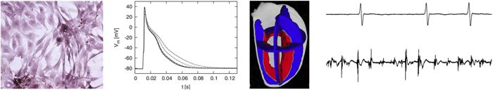
In this work, mathematical descriptions of the electrophysiology of cardiac fibroblasts will be developed and applied in computational simulations to gain insights into electrical interactions between fibroblasts and myocytes. A multi-domain model representing the electrical properties of cardiac tissue in different domains like myocytes, fibroblast and the extracellular space will be created. The fibroblast-myocyte ratio and the coupling will be varied to investigate the effects on conduction. Also the thickness of a fibroblast layer to build a barrier for conduction will be defined. Re-entry will be investigated in a 2D model and a schematic 2D description of infarct with islands of surviving myocytes will be developed. This will be used to validate if isolated myocytes in the vicinity of normal tissue might lead to so-called reflected re-entry. Additionally, the impact of fibrosis in generating so called complex fractionated atrial electrograms will be investigated.
Publications
- FB Sachse et al. A model of electrical conduction in cardiac tissue including fibroblasts. Ann Biomed Eng. (37), 874-889, 2009
- FB Sachse et al. Electrophysiological Modeling of Fibroblasts and Their Interaction with Myocytes. Ann Biomed Eng. (36), 41-56, 2008