Modelling of the human brain to predict spatial and temporal temperature profiles for the selective hypothermia treatment of an ischemic stroke
- contact:
In industrialized countries, stroke is the third-most widespread cause of death. Eighty percent of all strokes are caused by an ischemia resulting from a vascular obstruction, while intracerebral or subarachnoidal bleeding occurs in the remaining affected patients. The mortality rate of an ischemic stroke is about 25% while 35–55% of the patients experience of permanent disability.
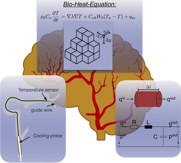
Hypothermia is a new, promising treatment of brain tissue after it has gone through reduced blood supply as observed in ischemic stroke patients. The treatment aims to ensure reduced metabolism and an anti-inflammatory effect to avoid possible severe brain damage. However, controlling the brain temperature and adjusting the cooling parameters are essential. Unfortunately, a selective, close-up measurement of the affected brain areas with intracranial probes is invasive and poses an additional risk of injury.
A new approach involves developing a procedure to cool the blood selectively in the proximity of the vascular obstruction combined with the reperfusion. The probe that is needed to cool the blood can be placed in blood vessels leading to the obstruction. The parenchym is perfused by capillary and is not accessible.
At the Institute of Biomedical Engineering in Karlsruhe, the main objective of this research project is the development of a brain model containing the most important blood vessels and blood-perfused brain tissue. The model contains the specific properties of the tissue (specific heat capacity and heat conduction) and takes the anatomy of the vessels into account to calculate the specific temperature profile. In ischemic stroke patients, the temperature in the particular brain areas depends on the severity of the vascular obstruction and the existence of collateral vessels. The model will be capable of predicting the spatial as well as temporal temperature profile in the parenchym. The model results will be compared with experimental results of in vitro measurements with simplified silicone or PVA brain models.